
2021 Contest Winners
Many of the contest participants used imaging techniques to capture beauty that normally goes unseen by the human eye, which as contest award winner Keunyoung Kim says, “can be so intricately and artistically organized.” In fact, Kim’s own work, “Mouse Retinal Ganglion Cells,” calls attention to the beauty of one of the “key components of visual information,” the retinal ganglion cell, which facilitates our ability to interpret the visual world.
By means of this capacity and the talents of these researcher-artists we can appreciate the tiny underwater organisms in Pichaya Lertvilai’s work, “Drifting World through the Scripps Plankton Camera,” cutting-edge nanobiology and nanotechnology research in Emil Karshalev’s “Vulture Perched on a Branch Waiting for its Next Meal” and Steven Edward Bopp’s “Polymer-encased Ag nanocube light source,” respectively; a pancreatic cancer tumor in Partha Ray’s image, “Red, White & Blue”; the common fruit fly transformed into a magnificent giant in Thomas Deerinck’s work, “Drosophila melanogaster”; and in Lorenzo Casalino’s work, “A Deceitful Handshake,” the microscopic giant that has dominated our lives over the last 15 months: COVID-19.
Finally, Eleanor Quirk’s image, “Diagonal Samples,” is accompanied by a thoughtful and passionate narrative that resets our lens to a broad perspective and to one of the core goals of the UC San Diego campus research mission: to help us understand the world we live in, empower us to better interact with our environments, and “ultimately make us better citizens of planet Earth.”
The Library’s Research Data Curation Program is grateful that despite these difficult, demanding and stressful times, the participants shared their work and gave us a glimpse into their worlds of research.
Judges' Award, Faculty/Scientist Participant Category
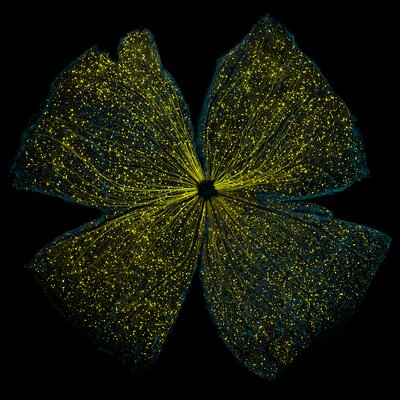
"Mouse Retinal Ganglion Cells" by Keunyoung Kim
Caption: Retinal ganglion cells in the whole-mounted mouse retina
Transduction of AAV2-GFP expression in the flat mounted mouse retina. Double labeling of Brn3a (blue) and GFP (yellow) in flat-mounted mouse retina. Note that GFP expression was observed in the somas and axons of RGCs. While working on a project focusing on the visual pathway, I tend to work with retinal ganglion cells quite often. This is because the retinal ganglion cell is one of the key components in the transportation of visual information from the eye to the brain. Imaging of retinal ganglion cells is an essential tool in measuring the level of damage sustained in the eye. I wanted to show the public that beauty lies not only in what is visible to the human eye but also in the unseen, particularly in the body’s central nervous system. I felt that this image captured the beauty of the retinal ganglion cells in whole-mount retina samples. It is amazing to see that microscopic structures that the naked eye cannot detect can be so intricately and artistically organized. The movement of connecting the world of microscopic science and the public is something I have always wanted to be a part of, and I feel that through UCSD library's research data curation program I was able to do so.
Keunyoung Kim is a Project Scientist associated with the National Center for Microscopy and Imaging Research at UC San Diego.
Judges' Award, Postdoctoral Participant Category
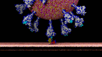
"A Deceitful Handshake" by Lorenzo Casalino
Caption: Ever wondered how SARS-CoV-2 latches onto human cells? Computer simulations capture the atomic-level details of this deadly encounter leading to infection.
The image shows the atom-by-atom reconstruction of the interaction between the severe acute respiratory syndrome coronavirus 2 (SARS-CoV-2, spherical particle coming from above) and the angiotensin-converting enzyme 2 (ACE-2, depicted with yellow and red cartoons) exposed on the host cell plasma membrane (flat lipid bilayer on the bottom). The virus utilizes a prominent glycoprotein, called spike (colored in gray), to latch onto ACE-2. This encounter complex allows the virus to gain entry into and infect the host cell, thus appearing like a “deceitful handshake”. At the Amaro lab, in the Chemistry and Biochemistry department at UCSD, we have been using biophysics and high-performance computing to fight COVID-19. As shown in this image, our effort culminated with the building of the all-atom SARS-CoV-2 viral envelope model, a system bearing over 300 million atoms integrating multiple cryoEM, cryoET, glycomic and lipidomic data. Molecular dynamics simulations of this system will usher in new opportunities for drug and vaccine design and enable the investigation of viral infection at subcellular scales. (References: Casalino L, Gaieb Z et al., Beyond Shielding: The Roles of Glycans in the SARS-CoV-2 Spike Protein. ACS Cent. Sci. 2020; Casalino L, Dommer AC, Gaieb Z, et al., AI-driven multiscale simulations illuminate mechanisms of SARS-CoV-2 spike dynamics. BioRxiv 2020).
Lorenzo Casalino is a postdoctoral researcher associated with Department of Chemistry and Biochemistry at UC San Diego and the Amaro Lab.
Judges' Award, Graduate Student Participant Category
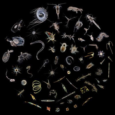
"Drifting World through the Scripps Plankton Camera" by Pichaya Lertvilai
Caption: The collage of tiny ocean drifters imaged by the Scripps Plankton Camera showing the hidden beauty of the underwater world that is unseen to naked eyes.
This image is a collage of many representative species of plankton from the Scripps Plankton Camera, showing the hidden beauty of these tiny organisms that cannot be seen by naked eyes. The Scripps Plankton Camera, developed by the Jaffe Lab, is an underwater microscope system that autonomously collects in situ images of plankton at Scripps Pier. The system utilizes darkfield microscopy technique to image these small and mostly transparent marine organisms. The underwater microscope has been running for more than five years and has collected more than millions of images of these plankton. The Jaffe Lab collaborates with many ecologists, both at UCSD and at other institutions around the world, to capitalize on this novel underwater imaging technology and on the large dataset it produces. The system has helped scientists better understand these organisms and can ultimately contribute to new discoveries of the ecology of the ocean.
Pichaya Lertvilai is a graduate student associated with the Scripps Institution of Oceanography and the Jaffe Laboratory of Underwater Imaging.
Judges' Award, Undergraduate Participant Category
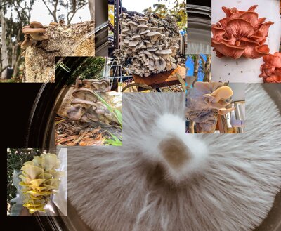
"Hyphi" by Nika Redburn and Will Tanaka
Caption: The Web of Life
The background photo is of a vigorous edible mushroom culture on a agar petri dish made in a student's dorm. The surrounding photos are different types of mushrooms that were grown by the same student using waste such as coffee grounds and tea leaves collected from the campus. The mushrooms were grown at Roger's Urban Farm, a student-run community garden on the UCSD campus.
Nika Redburn and Will Tanaka are undergraduate students associated with the Department of Nanoengineering and the Department of Biological Sciences (Neurobiology) at UC San Diego, respectively, and the Roger's Urban Farmlab (RUF).
Open Voting Winner
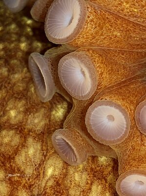
"Bigeye Octopus Suckers" by Adi Khen
Caption: A sucker for suckers
Closeup shot of the suckers of a North Pacific Bigeye octopus ("Octopus californicus"), collected from a deep-sea trawl on a research vessel and kept in Scripps Institution of Oceanography's Experimental Aquarium.
Adi Khen is a graduate student associated with the Marine Biology Research Division (MBRD) at Scripps Institution of Oceanography and the Smith Lab.
Judges' Award Winner, Honorable Mention
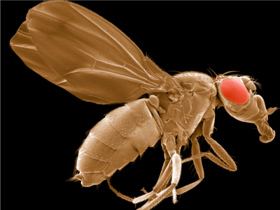
"Workhorse of genetics" by Thomas Deerinck
Drosophila melanogaster
Long considered the linchpin of genomics research since they are easy to manipulate, inexpensive to maintain, have short generation times and share many genes involved in human diseases, these model organisms have led to countless discoveries in biomedicine. This scanning electron micrograph was generated as part of a collaboration between the National Center for Microscopy and Imaging Research at UCSD and the laboratory of renowned UCSD scientist Dr. Michael Karin to better understand genes and aging (see: Lee et al., Science. 2010 Mar 5;327(5970):1223-8).
Thomas Deerinck is a faculty member associated with the National Center for Microscopy and Imaging Research at UC San Diego.
Judges' Award Winner, Honorable Mention
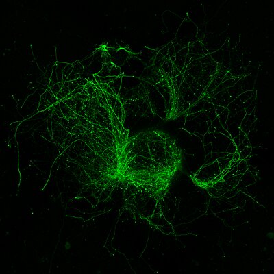
"Whirlwind of neurites extending from fluorescent neurons in cultured explants of embryonic mouse brain tissue" by Jess Du
Molecular programs for neuronal development can unfold semi-autonomously, allowing these neurons from tiny pieces of embryonic brain tissue on a vast, alien culture dish to grow into elaborate forms
Embryonic mouse neurons labeled with green fluorescent protein, with profusions of neurites extending outwards in swirls. Image captured on a confocal STORM microscope at 10x magnification. The neurons are nascent inhibitory interneurons within tissue explants microdissected from embryonic mouse brains and grown on top of a primary cortical culture for 8 days.
Jess Du is a graduate student associated with the Department of Neurosciences at the UC San Diego School of Medicine and The Lippi Lab
Judges' Award Winner, Honorable Mention
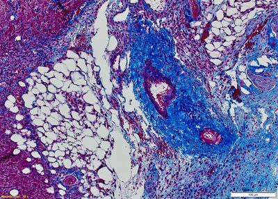
"Red, White & Blue" by Partha Ray
Trichrome staining was performed on a Formalin-Fixed Paraffin-Embedded (FFPE) mouse Pancreatic Cancer Tumor slide. The muscle tissues and collagens differentially stain in Red and Blue color, respectively
Formalin-Fixed, Paraffin-Embedded (FFPE) tissue slides from a mouse pancreatic cancer tumor were stained with the Trichrome histological staining method. Collagens and muscle tissues are stained differentially in blue and red color, respectively.
Partha Ray is a faculty member associated with the Division of Surgical Oncology, Department of Surgery, Moores Cancer Center at the UC San Diego School of Medicine.