
2023 Contest Winners
For the 2021 and 2022 contests, we invited campus researchers to submit images related to their research. This year we asked the UC San Diego and San Diego communities to vote on images deposited in the Library’s Research Data Repository.
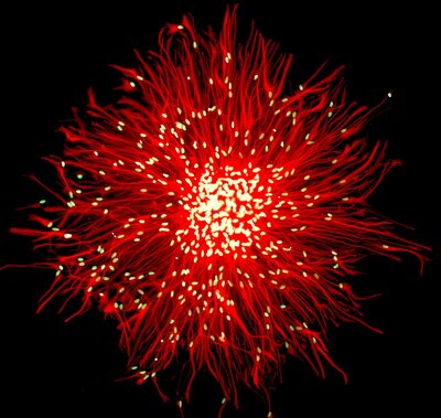
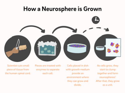
"Human Spinal Cord Neurosphere" by Michael Weible
Tiny stem cell clusters hold big promise for medical science
A human spinal cord neurosphere is a fascinating structure that plays a crucial role in our nervous system. Imagine a tiny, round cluster of cells that contains specialized cells called neural stem cells. These neural stem cells have the remarkable ability to self-renew and differentiate into various types of nerve cells found in the spinal cord. The spinal cord neurosphere acts as a miniature laboratory where scientists can study and understand how our nervous system develops and functions. It holds great potential for research in treating spinal cord injuries and neurological disorders.
By studying the neurosphere, scientists hope to unlock the secrets of nerve cell regeneration and develop new therapies to help people with spinal cord injuries regain mobility and function.
How incredible that something so small can hold so much promise for advancing medical science!
To learn more about this image, visit the Library's Research Data Repository by clicking here.
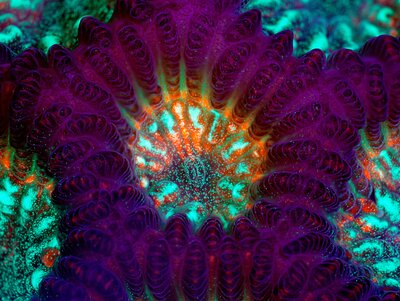
"Live Green Brain Coral (Genus Goniastrea)" by James Nicholson
LED lights shine a spotlight on hard-to-see coral anatomy
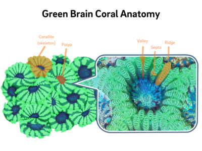
Most corals are made up of many tiny animals called polyps, living together in a colony. In this image, you can see one full polyp in the center with four others surrounding it. The purple ring is its corallite, or skeleton.
Why does the coral’s skeleton look purple in the image but not in the illustrations? Scientists used an LED light to illuminate these coral parts in color, making them easier to observe and study, and quite pleasing to the eye.
More about brain coral:
Corallite (skeleton): The corallite is where the polyps of the coral attach themselves and build the coral structure.
Polyp: Imagine a tiny, colorful sea creature with a soft body. This creature is called a polyp. Each polyp is responsible for secreting calcium carbonate and creating the coral structure. They have tentacles that help them catch food and defend themselves.
Valley: A valley in brain coral refers to the grooves or channels that run between the ridges.
Septa: The septa in brain coral have a pattern of ridges and grooves and help provide support and structure to the coral.
Ridge: The ridges give the brain coral its distinct texture and help protect the delicate polyps inside.
To learn more about this image, visit the Library's Research Data Repository by clicking here.
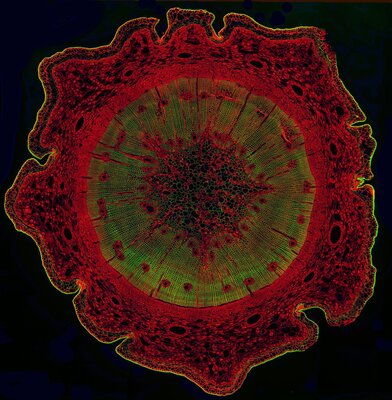
"Pine Tree Stem (Genus Pinus)" by Lauren Piedmont
Fluorescent images give scientists a closer look at the details of a pine tree stem cross-section
This image of a pine tree cross-section was taken with a microscope. Fluorescent lights are shone on the specimen, resulting in the bright colors you see. This technique—called fluorescent microscopy—helps scientists (and us) see tiny details of the tree’s stem.
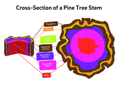
More about the pine tree stem cross-section:
Periderm: the protective outer layer of the stem, replacing the epidermis in older parts of the tree. It consists of several distinct layers, including the cork cambium, cork cells, and phelloderm. The periderm acts as a barrier against external damage, pathogens, and water loss.
Xylem and Phloem: xylem tissue is responsible for transporting water and nutrients from the roots to other parts of the tree, while phloem tissue transports sugars produced during photosynthesis to various parts of the tree.
Vascular Cambium: a thin layer of cells located between the xylem and phloem. It is responsible for secondary growth in the stem, resulting in the thickening of the tree over time. The vascular cambium produces new xylem cells towards the inside of the stem and new phloem cells towards the outside, contributing to the tree's expanding girth.
Pith: at the very center of the cross-section, there is often a central, cylindrical region known as the pith. The pith consists of soft, spongy tissue and serves as a storage area for nutrients.
To learn more about this image, visit the Library's Research Data Repository by clicking here.
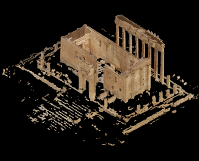
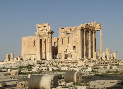
"Temple of Bel in Palmyra, Syria" by Scott McAvoy
Thousands of crowdsourced images allow scientists to digitally recreate this ancient temple
Constructed over the first and second centuries, this temple was a center for religious life in the ancient city of Palmyra. But in 2015, it was destroyed.
Now, the temple has been digitally reconstructed using cutting-edge, three-dimensional modeling and artificial intelligence to stitch together crowdsourced photographs and recreate the structure.
The project has helped digitally preserve more than a dozen lost reliefs, sculptures, frescoes and paintings, all publicly available on the UC San Diego Library’s Digital Collections website.
To learn more about this image, visit the Library's Research Data Repository by clicking here.
Locate Palmyra, Syria in Google Earth by clicking here.
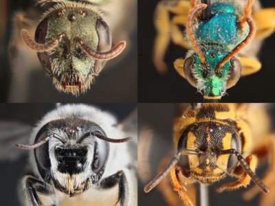
"Face the Bees" by Keng-Lou James Hung and Susan Chen
Before we ask how to save the bees, we must know which bees to save
Surveys revealed that San Diego County is home to roughly 700 species of native bees, some of which were previously unknown to science. Now that scientists know what these bees are and where they live, they can take steps to protect their populations.
Pictured (clockwise from top left): brown-bellied metallic sweat bee (Lasioglossum brunneiventre), Texas striped sweat bee (Agapostemon texanus), wide-belted dark bee (Stelislaticincta), orange-tipped woodborer bee (Lithurgopsis apicalis).
To learn more about this image, visit the Library's Research Data Repository by clicking here.
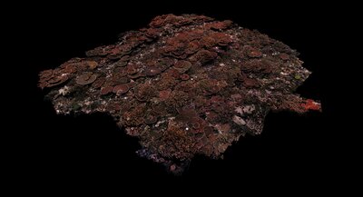
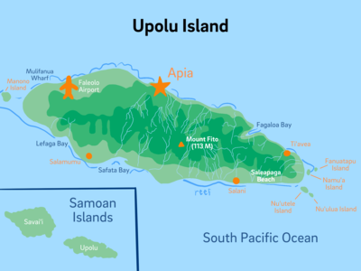
"Coral Reef of Samoa" by Sandin Lab, Scripps Institution of Oceanography, UC San Diego
Digital models like the one seen here help scientists study coral reefs
Imagine if you could see a coral reef without the ocean water in the way. By taking thousands of photographs and stitching the images together, researchers at Scripps Institution of Oceanography create realistic models of underwater habitats. This is a tennis court-sized section of coral reef from the island of Upolu in Samoa. Look at the large corals that look like tabletops. This coral reef is thriving!
To learn more about this image, visit the Library's Research Data Repository by clicking here.
Locate Upolu, Somoa in Google Earth by clicking here.
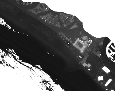
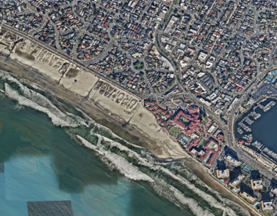
"Digital Map of Coronado Beach" by Adam P. Young, Reinhard E. Flick, Timu W. Gallien, Sarah N. Giddings, R.T. Guza, M. Harvey, Luc Lenain, B.C. Ludka, W. Kendall Melville, and W.C. O'Reilly
Elevation map aids the study of shoreline changes in Coronado, California during El Niño
This image shows an elevation map of Coronado Beach made using lasers collected from an airplane in 2016 to study the impacts of El Niño along the Southern California coast. The 2015-16 El Niño was one of the strongest recorded El Niño events and generated record high water levels. Researchers used this map to measure changes along the coast and found that although some areas experienced significant change, many areas in Southern California were sheltered from damage.
About El Niño:
El Niño is a weather phenomenon that occurs irregularly in the tropical Pacific Ocean. It is characterized by a warming of the ocean surface temperatures, specifically in the central and eastern regions of the Pacific Ocean. El Niño events typically last for several months to a few years and can have significant impacts on weather patterns around the world.
During a typical El Niño event, the trade winds (prevailing winds blowing from east to west) weaken or even reverse direction, causing the warm surface waters to move toward the eastern Pacific. This movement of warm water alters the normal patterns of atmospheric circulation, resulting in changes to global weather patterns.
To learn more about this image, visit the Library's Research Data Repository by clicking here.
Locate Coronado Beach, California in Google Earth by clicking here.
Images from the Neolithic Site of Çatalhöyük
Çatalhöyük (pronounced "cha-tal hay OOK") is highly regarded as one of the most important archaeological discoveries in the world. It dates back to the Neolithic period, around 7100-5900 B.C.E.
Pictured (clockwise from top left):
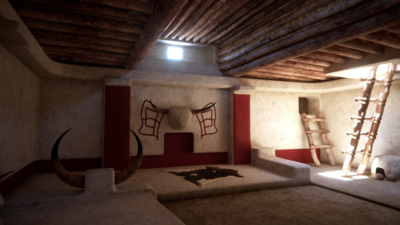
Image 1: "Çatalhöyük, South Area, Shrine" by Nicola Lercari, Gesualdo Busacca, Grant Cox, Jad Aboulhosn, Anaïs Guillem, and Arianna Campiani
Three-dimensional reconstruction of the ancient archaeological site Çatalhöyük located in present day Turkey
The decoration of this building was dominated by cattle heads located in the central part of the eastern and northern walls.
To learn more about this image, visit the Library's Research Data Repository by clicking here.
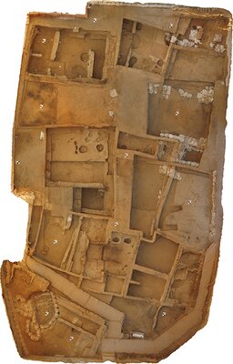
Image 2: "Çatalhöyük, North Area" by Arianna Campiani, Nicola Lercari, Ashley Lingle, Moataz Dahabra, Manuel Dueñas García, Estrella García, Tristan Yang, John Flynn, and Christopher Reps
A laser scan of Çatalhöyük helps map out the archaeological site
Scientists used a special laser scanning technique called “area-wide terrestrial laser scanning” to gather detailed information about the land and structures of Çatalhöyük.
To learn more about this image, visit the Library's Research Data Repository by clicking here.
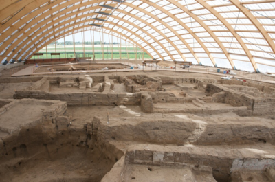
Image 3: "Neolithic Site of Çatalhöyük" (2019) by Jason Quinlan
Locate Çatalhöyük in the Konya plain, near the town of Çumra in Southern Anatolia, Turkey in Google Earth by clicking here.