
Honorable Mentions
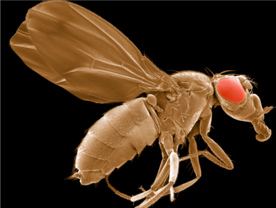
"Workhorse of genetics" by Thomas Deerinck
Drosophila melanogaster
Long considered the linchpin of genomics research since they are easy to manipulate, inexpensive to maintain, have short generation times and share many genes involved in human diseases, these model organisms have led to countless discoveries in biomedicine. This scanning electron micrograph was generated as part of a collaboration between the National Center for Microscopy and Imaging Research at UCSD and the laboratory of renowned UCSD scientist Dr. Michael Karin to better understand genes and aging (see: Lee et al., Science. 2010 Mar 5;327(5970):1223-8).
Thomas Deerinck is a faculty member associated with the National Center for Microscopy and Imaging Research at UC San Diego.
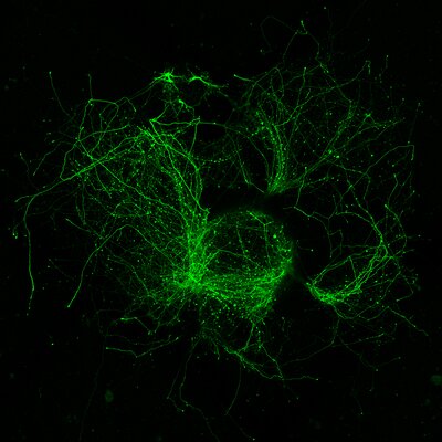
"Whirlwind of neurites extending from fluorescent neurons in cultured explants of embryonic mouse brain tissue" by Jess Du
Molecular programs for neuronal development can unfold semi-autonomously, allowing these neurons from tiny pieces of embryonic brain tissue on a vast, alien culture dish to grow into elaborate forms
Embryonic mouse neurons labeled with green fluorescent protein, with profusions of neurites extending outwards in swirls. Image captured on a confocal STORM microscope at 10x magnification. The neurons are nascent inhibitory interneurons within tissue explants microdissected from embryonic mouse brains and grown on top of a primary cortical culture for 8 days.
Jess Du is a graduate student associated with the Department of Neurosciences at the UC San Diego School of Medicine and The Lippi Lab
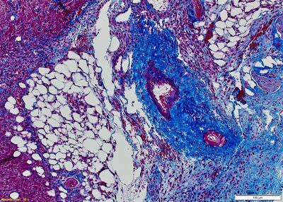
"Red, White & Blue" by Partha Ray
Trichrome staining was performed on a Formalin-Fixed Paraffin-Embedded (FFPE) mouse Pancreatic Cancer Tumor slide. The muscle tissues and collagens differentially stain in Red and Blue color, respectively
Formalin-Fixed, Paraffin-Embedded (FFPE) tissue slides from a mouse pancreatic cancer tumor were stained with the Trichrome histological staining method. Collagens and muscle tissues are stained differentially in blue and red color, respectively.
Partha Ray is a faculty member associated with the Division of Surgical Oncology, Department of Surgery, Moores Cancer Center at the UC San Diego School of Medicine.